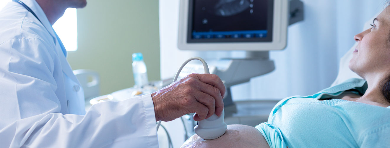
Ultrasound is an imaging test that allows a healthcare provider to see inside the body. It uses high-frequency sound waves to create on-the-spot pictures or video of internal organs or other tissues. Such pictures are called sonograms. An experienced doctor (called an ultrasound technologist or sonographer) performs the procedure.
Dr. Yugal Goyal is a trusted sonography doctor in Siliguri. With an experience spanning years, he is proficient in providing safe and precise ultrasound diagnostics. He uses advanced machines and technology to perform this procedure, delivering precise reports.
The cost of an ultrasound in Siliguri is between ₹500-₹3500. However, the actual costs depend on the type of ultrasound service, the purpose of the scan, Doppler requirement, and 2D,3D,4D imaging, etc. Here is a breakdown of the costs of ultrasound -
| Type of Ultrasound | Approximate Cost in Siliguri |
|---|---|
| Pregnancy USG | ₹500 -₹1200 |
| Whole abdomen USG | ₹650-₹1500 |
| Upper abdomen | ₹1050-₹1200 |
| Lower abdomen | ₹1050-₹1200 |
| KUB + Pelvis | ₹1050-₹1200 |
| Foetal Doppler | ₹2200 |
| Anomaly/Foetal scan | ₹3300 |
Disclaimer - The above costs are indicative and not fixed. Actual charges may vary depending on the factors mentioned above. Please confirm the current price with Dr. Yugal Goyal's diagnostic centre before undergoing the scan.

The types of ultrasound scans available at Dr. Goyal's Diagnostic Centre are abdominal, pelvic, obstetrics ( pregnancy ), thyroid, breast, Doppler, and others. The type of ultrasound depends on a patient's symptoms, medical history, and clinical requirements. A trained ultrasound professional can determine which scan is appropriate and interpret the findings after considering the patient's overall health.
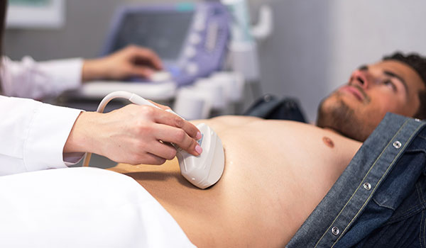
It is performed to obtain images of the liver, gallbladder, pancreas, spleen, kidneys, and urinary bladder as prescribed by the doctor. Being one of the most common ultrasound scans in Siliguri, it is usually prescribed when a patient has symptoms such as abdominal pain, inexplicable discomfort, jaundice, or changes involving the abdominal region.
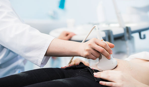
It examines structures located in the pelvic region for both men and women. In females, it is done to provide images of the uterus, cervix, ovaries, and the neighbouring pelvic regions. In males, it is done to obtain images of the prostate gland, urinary bladder, seminal vesicles, etc.
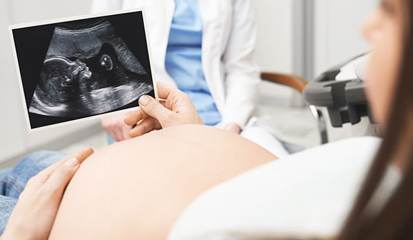
It provides information about fetal growth, fetal position, heartbeat, placental location, amniotic fluid, and more. It is done during different stages of pregnancy to either assess early pregnancy stages or fetal growth and development during the later stages.
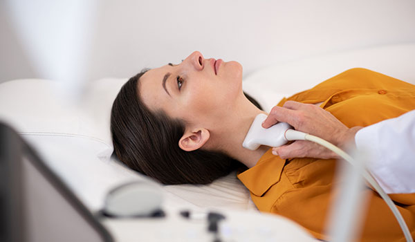
It is recommended to evaluate the thyroid gland and the tissues surrounding it. It is prescribed when there is a thyroid nodule, swelling in the neck, an enlarged thyroid gland, or other suspected abnormality.
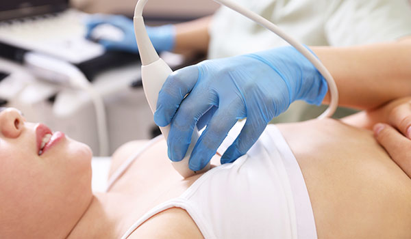
This examination is done to examine a breast lump, localised pain, swelling, or other similar area of concern. It helps distinguish between certain fluid-filled structures such as cysts and the solid areas within the breast.
It is a specialised form of ultrasound that evaluates blood flow through blood vessels. It is different from traditional ultrasound that creates images of anatomical structures, while Doppler provides information about the movement of blood, its direction, and the characteristics of blood flow.
An ultrasound may be required when a doctor needs more information about certain symptoms such as a lump, swelling, abdominal or pelvic discomfort, to examine the condition of the foetus during pregnancy, or to evaluate internal organs like the kidneys, urinary bladder, thyroid, uterus, ovaries, etc. Here is a list of some common situations in which a doctor might recommend an ultrasound scan in Siliguri -
A patient might require an ultrasound when they are experiencing some symptoms that do not have a clear explanation, such as abdominal or pelvic pain, swelling, a lump, discomfort, or changes in a particular part of the body.
A doctor may recommend an ultrasound scan to check whether a finding has changed in size or appearance or to verify how a condition is progressing.
Pregnant women might be prescribed an ultrasound at different stages of their pregnancy to monitor foetal growth, development, position, heartbeat, placental location, or some other pregnancy-related findings.
Depending on the symptoms and clinical requirement, a patient might require an image of the liver, gallbladder, kidneys, urinary bladder, thyroid, uterus, ovaries, breasts, or more. It can help identify structural changes, cysts, stones, fluid collections, nodules, and other findings.
When a doctor needs to check blood circulation in specific blood vessels, the patient may need an ultrasound. Doppler techniques provide information about the movement and direction of blood flow.
Preparing for an ultrasound involves fasting and having a full bladder. However, both are optional and need to be followed only when instructed. Generally, preparation for an ultrasound scan in Siliguri is fairly simple and may vary depending on the type of scan, the body area being examined, and the purpose of the examination.
For some abdominal ultrasound scans, patients may be asked to avoid food and drinks for a specific period before the scan. This is recommended to reduce the gas in the digestive tract so that structures like the gallbladder and abdominal organs can be examined more clearly.
For certain pelvic and urinary system exams, patients may be asked to arrive with a full bladder. This is recommended because it provides a clear image of the pelvic structures. Thus, patients should drink a specified amount of water and avoid urinating until the scan is over.
It depends on the stage of pregnancy and the type of examination. Some early pregnancy or pelvic examinations may require a full bladder, while most later pregnancy scans may not.
Dr. Yugal Goyal is a consultant radiologist based in Siliguri. With extensive experience in radiology and diagnostic imaging, he is known for providing the best ultrasound scan in Siliguri. Here are some more reasons why he is the first choice among patients -
Dr. Goyal holds an MBBS and an MD in Radiodiagnosis, along with years of experience in ultrasound diagnostics. This bolsters patients' trust in his medical acumen.
He is one of the very few doctors in Siliguri to provide the services of tele-ultrasound for remote interpretation and reporting of medical images. His longstanding experience with digital radiology workflows, PACS, and advanced imaging supports healthcare providers beyond his local clinic.
Dr. Goyal has experience in interpreting a wide spectrum of imaging, including X-rays, ultrasound, advanced CT, and MRI examinations. His expertise includes CT angiography, CT perfusion, MR angiography, MR perfusion, and MR spectroscopy.
He has extensive experience in USG and guided biopsies, FNAC, and percutaneous drainage, nephrostomy, biliary drainage, PTBD stenting, radiofrequency ablation, and microwave ablation. This enables him to diagnose complex issues accurately.
Yes, sometimes you can eat before an ultrasound. However, eating or not eating before an ultrasound depends on the type of ultrasound examination being undertaken. For an abdominal ultrasound, you may be asked to fast for several hours before the examination. This is because food and gas can affect the visibility of some abdominal organs. For a pelvic ultrasound, you may be asked to keep your bladder full. For a pregnancy ultrasound, preparation depends on the type of scan. For thyroid or breast ultrasounds, fasting is usually not required.
The amount of water to drink depends on the type of scan and the instructions provided by the diagnostic centre. For pelvic or lower abdominal ultrasound, generally 500-750 ml of water one hour before is advised. KUB ( Kidney, Ureters and Bladder) and bladder ultrasound, it is advisable to keep a full bladder. For early pregnancy ultrasound, a full bladder is sometimes recommended. However, for later pregnancy scans, a full bladder is not generally required.
The right time to get an ultrasound depends on your symptoms, medical condition, pregnancy status, and the type of examination required. Generally, the doctor recommends an ultrasound scan if you have inexplicable pain or discomfort, for pregnancy assessment, to monitor an existing condition such as in the liver, gallbladder, kidney, thyroid, etc., to assess blood flow, or if you have unusual swelling, a lump, or changes in the abdomen.
Ultrasound and sonography refer to the same type of medical imaging. Both involve the use of high-frequency sound waves to create images of structures inside the body. However, there is a minor difference between the two. Ultrasound refers to the imaging technology or the examination itself. Sonography means the process of creating and examining images using ultrasound waves.
A USG, or ultrasonography, is an imaging examination that uses high-frequency sound waves to create images of organs, tissues, blood vessels, and other structures inside the body. A doctor may recommend a USG test in Siliguri for several reasons, such as to investigate unexplained abdominal or pelvic pain, swelling, etc.; to examine internal organs like the gallbladder, liver, kidney, ureters, etc.; to monitor pregnancy; to evaluate blood flow; to inspect a lump or swelling; or to assess changes over time.
Preparation for an ultrasound scan depends on the type of ultrasound being performed. Some scans may need specific preparation, while others may not. For an abdominal ultrasound, you may be asked to avoid eating or drinking for a few hours before the scan. This is to reduce gas, which can become an obstruction to clear images. For a pelvic ultrasound, it is advisable to keep a full bladder to get clear images. For a KUB Kidney, Ureter, and Bladder) ultrasound, a full bladder may be required depending on the examination. For a pregnancy ultrasound, preparation varies according to the stage of pregnancy and the type of scan. However, thyroid, breast, and other similar scans do not require any special preparation.
The time required for an ultrasound depends on the type of examination, the area to be examined, and the level of detail required by the doctor. A simple scan that includes a single area and does not require too many details may be completed within a short time. However, a more detailed scan or one involving many areas may require a longer time.
Yes, ultrasound is generally considered a safe imaging technique for both children and adults when it is performed appropriately. Unlike X-Rays and CT scans, conventional ultrasound uses high-frequency sound waves rather than ionizing radiation to produce images. It may be used in children and adults to examine organs, soft tissues, blood vessels, or any other area of concern.
Most ultrasound examinations do not cause pain or extreme discomfort. During the procedure, a healthcare professional applies a small amount of gel to the skin and moves a device called a transducer over the area to be examined. However, some patients may feel mild pressure or temporary discomfort when the transducer is moved over the area that is already tender, swollen, or painful. This discomfort does not last long and goes away quickly after the examination.
What Patients Say
Clear & Reassuring
The entire process was explained clearly, and I felt comfortable asking questions. The consultation was professional, calm and reassuring.

Amit Sharma, Siliguri
Professional Experience
Very professional approach and detailed attention to the case. I appreciated how patiently everything was explained before proceeding.

Priya Das, Siliguri
Thorough Consultation
Dr. Goyal took the time to understand the concern and explain the findings in simple language. A very reassuring experience.

Rahul Agarwal, Jalpaiguri
Patient & Approachable
The consultation was handled with patience and care. Dr. Goyal explained the medical details in a way that was easy to understand.

Sneha Roy, Malbazar
Sanjay Gupta
From the initial consultation to understanding the imaging findings, everything was handled in a clear and professional manner.

Smooth Experience, Siliguri
Copyrights © 2026, Dr. Yugal Goyal.
