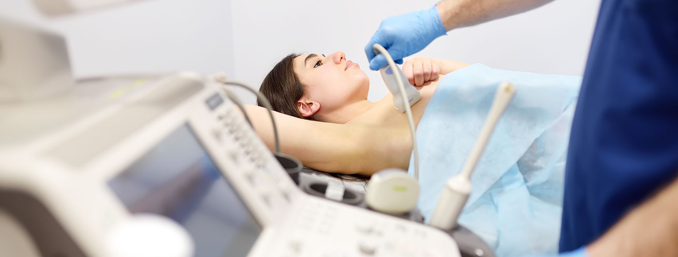
Breast ultrasound is a non-invasive imaging examination that uses high-frequency sound waves to create detailed images of the breast tissue. It is used to evaluate a breast lump or other abnormality found during a medical check-up or some other imaging test. Unlike mammography, ultrasound does not use ionizing radiation.
For patients looking for breast ultrasound scans in Siliguri, Dr. Yugal Goyal provides the best sonography services. The ultrasound is non-invasive and generally painless. It does not require needles or injections and does not use ionizing radiation.
According to John Hopkins Medicine, a breast ultrasound is an imaging examination that uses sound waves to look inside the breasts. It can help your healthcare provider to detect breast problems. The healthcare provider can also see how well blood is flowing to areas in the breast. This examination is generally recommended when a change has been seen in a mammogram or when a change is felt but is not reflected in the mammogram.
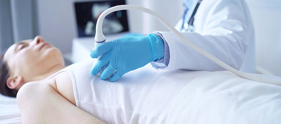
To perform a breast ultrasound, a device called a transducer is moved over the skin after applying ultrasound gel. The sound waves reflected from the tissues are converted into images that can be viewed on a monitor.For those seeking breast USG test in Siliguri, this non-invasive examination can help evaluate breast changes and abnormalities.
A doctor may recommend a breast ultrasound when there is a specific area of concern that requires further evaluation. It is often used to investigate findings that are detected during a clinical assessment or mammography or MRI. Some other reasons why patients may need a breast scan in Siliguri are -

a new lump or some other palpable area of concern may require imaging. Ultrasound can help determine whether the finding appears to be a cyst, a solid mass, or another type of tissue abnormality.

if a mammogram shows an area that requires closer assessment, ultrasound can be recommended to determine whether the finding appears to be a cyst, a solid mass, or some other tissue abnormality.

when a patient is experiencing breast pain or some other localised breast symptoms, an ultrasound may help in determining its cause.

ultrasound may also be used for clinical concerns such as nipple discharge or some changes in the breast skin.
dense breast tissue can make some abnormalities more difficult to see on the mammogram. In selected patients, ultrasound can be used as an additional imaging test. However, ultrasound does not replace mammography for breast cancer screening.

Breast ultrasound can provide detailed information about the breast tissue. It helps distinguish between fluid-filled structures, solid masses, and other changes in the breast tissue. Depending on the clinical findings, age, symptoms, and reason for the examination, a doctor may recommend a breast USG test in Siliguri for the following reasons -
it can identify fluid-filled structures within the breast. It is particularly useful for distinguishing a fluid-filled cyst from a solid mass.
a solid area can be identified and evaluated based on its size, shape, margins, internal appearance, etc.
some breast abnormalities contain both fluid-filled and solid components. Ultrasound can reflect these different components and provide information for further assessment.
small or localised abnormalities that may not be easily characterised in a medical checkup can be examined more clearly with an ultrasound.
ultrasound can sometimes identify lesions that are difficult to visualise on mammography because of dense breast tissue. However, it cannot be considered a replacement for mammography.
The key difference between a breast ultrasound and mammography lies in the technologies that they use. Breast ultrasound uses sound waves to create images, while mammography uses low-dose X-rays. The choice between the two depends on various factors such as age, symptoms, breast density, clinical findings, pregnancy status, etc.
| Feature | Breast Ultrasound | Mammography |
|---|---|---|
| Imaging method | Uses high-frequency sound waves | Uses low-dose X-rays |
| Radiation Exposure | No ionizing radiation | Uses a small amount of ionizing radiation |
| Best known for | Evaluating lumps and distinguishing cystic from solid lesions | Breast cancer screening and detecting certain abnormalities including calcifications |
| Breast Lumps | Determines whether a lump is fluid-filled or solid | Can evaluate many breast abnormalities along with other imaging tests |
| Pregnancy | Can be used for breast imaging | Avoided unless specifically needed because it uses X-rays |
| Screening Role | Not a replacement for mammography | Key test for breast cancer screening |
Note: The above prices are indicative and may vary between diagnostic centres and according to the examination required. The final cost should be confirmed with the diagnostic centre before the examination.
A breast ultrasound is a simple examination. The patient is asked to lie on their back during the examination. She may be asked to keep one arm above her head to provide better access to the breast.
An ultrasound gel is applied to the skin. The ultrasound professional or radiologist then moves the transducer over the breast to capture images from different angles. In case there is a specific lump, area of pain, or some other concern, then particular attention may be paid to that region.
After the scan, the images are reviewed, and a report of the ultrasound is prepared. The examination does not involve radiation, needles, or injection. The patient may feel some pressure when the transducer is moved across the breast, particularly over a tender area.
A breast ultrasound requires little or no preparation at all. The patient can normally eat and drink unless the healthcare provider gives special instructions. For a breast USG test in Siliguri, a patient can -
A previous ultrasound report can be useful in comparison with the current examination. This can help the doctor in determining whether the finding is new, has remained stable, or is changing.

The cost of breast USG scan in Siliguri varies depending on factors such as the type of examination, the diagnostic centre, reporting equipment, and whether any additional imaging is included. A list of prices for different breast ultrasound services is given below -
| Breast Ultrasound Service | Indicative Cost in Siliguri |
|---|---|
| Breast Ultrasound | ₹800-₹1500 |
| Bilateral Breast Ultrasound | ₹1000-₹1800 |
| Breast Ultrasound with additional targeted evaluation | ₹1000-₹2000 |
Note: Bilateral breast ultrasound is an ultrasound that examines both breasts.
Disclaimer: These are indicative prices and may vary depending on the diagnostic centre, examination requirements, and any additional procedure or laboratory charges. Patients should confirm the current cost with the centre before scheduling the examination.
While looking for breast ultrasound, patients may want to go for quick and clear images with compassionate care. All these features are provided at Dr.Yugal Goyal's Diagnostic Centre. With advanced technology and a patient-centric approach, he is one of the most trusted doctors when it comes to breast ultrasound in Siliguri. Here are some key reasons to choose him -
he holds an MBBS and an MD in Radiodiagnosis, along with longstanding experience in Radiology. Thus, he is adept at handling complex medical cases with ease.
He is one of the very few doctors in Siliguri to provide tele-ultrasound services. This will help long-distance patients in obtaining his expert diagnostic imaging services.
He offers a wide spectrum of image-guided procedures, including FNAC, percutaneous drainage, nephrostomy, biliary drainage, and PTBD stenting. Thus, his diagnostic centre is a one-stop solution for medical issues requiring various solutions.
His ultrasound centre offers a range of imaging services including CT scans, MRI, Ultrasound, X-Rays, CT angiography, CT perfusion, MR angiography, MR perfusion, and MR spectroscopy.
A breast ultrasound is a non-invasive imaging test that uses high-frequency sound waves to create detailed images of the breast. Unlike mammography, it does not use ionizing radiation. It is commonly used to evaluate a breast lump or other abnormality, determining whether an area appears fluid-filled, solid, or mixed. It may also be recommended to further assess a finding obtained from a mammogram or during another clinical breast examination.
Yes, a breast ultrasound is usually considered safe. This is because it uses high-frequency sound waves, which are non-invasive, to create images of breast tissue. Thus, it can be used even during pregnancy or breastfeeding.
Breast ultrasound is painless and does not involve needles, injection, or radiation. The patient may feel mild pressure or discomfort as the transducer is moved across the breast. However, the discomfort usually goes away as soon as the examination is done.
A breast ultrasound in Siliguri usually takes around 15-30 minutes. However, the exact time can vary depending on the area being examined and whether additional images are needed. After the scan, patients can return to their normal activities immediately after the scan.
No, fasting is generally not required before a breast ultrasound. The patient can eat, drink water, and take regular medications as advised before the examination.
Yes, breast ultrasound can detect and evaluate many breast lumps or masses. It can provide detailed images of the area and help determine whether a lump is fluid-filled, solid, or mixed. It can also evaluate its size, location, shape, and margins. This helps the radiologist decide whether further evaluation is needed.
No, breast ultrasound generally cannot replace mammography. This is because mammography is an important imaging tool for breast cancer screening, including detection of calcifications, that may not be visible on an ultrasound. On the other hand, breast ultrasound is useful for evaluating a breast lump and determining whether the lump is fluid filled or solid, or mixed.
Yes, breast ultrasound can generally be performed during pregnancy. This is because it uses sound waves rather than ionizing radiation. Thus, it is a non-invasive procedure, making it a useful imaging option to assess a breast lump or other breast changes during pregnancy.
What Patients Say
Clear & Reassuring
The entire process was explained clearly, and I felt comfortable asking questions. The consultation was professional, calm and reassuring.

Amit Sharma, Siliguri
Professional Experience
Very professional approach and detailed attention to the case. I appreciated how patiently everything was explained before proceeding.

Priya Das, Siliguri
Thorough Consultation
Dr. Goyal took the time to understand the concern and explain the findings in simple language. A very reassuring experience.

Rahul Agarwal, Jalpaiguri
Patient & Approachable
The consultation was handled with patience and care. Dr. Goyal explained the medical details in a way that was easy to understand.

Sneha Roy, Malbazar
Sanjay Gupta
From the initial consultation to understanding the imaging findings, everything was handled in a clear and professional manner.

Smooth Experience, Siliguri
Copyrights © 2026, Dr. Yugal Goyal.
