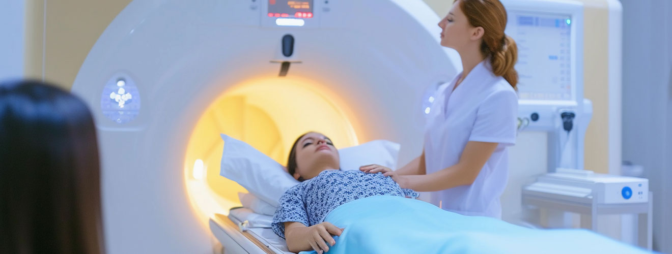
MRI is an imaging technique that uses a magnetic field and computer-generated radio waves to create clear and detailed images of the organs and tissues of the body, particularly the soft tissues. The soft tissues include the brain, spinal cord, muscles, joints, ligaments, and organs.
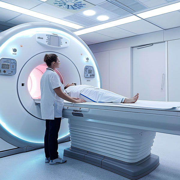
An MRI test in Siliguri involves an MRI scanner, which is a large, tunnel-shaped machine that is open from the front and back and closed from the sides and the top. An MRI is generally safe, and unlike X-rays and CT scans, it does not use ionising radiation. Instead, it uses a strong magnetic field that is harmless. However, it can affect implanted medical devices and metal objects.
An MRI is recommended when detailed imaging is needed to investigate symptoms, identify abnormalities, examine an injury, or monitor an existing condition. MRIs are particularly useful for examining soft tissues such as the brain, spinal cord, muscles, ligaments, etc. They can also provide information about blood vessels and the biliary system. MRI imaging in Siliguri is recommended for the following reasons –
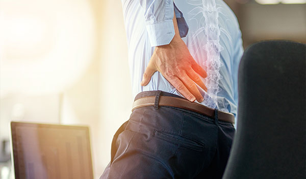
it helps doctors investigate probable causes of symptoms such as persistent headaches, back pain, joint pain, numbness, weakness, or other issues.
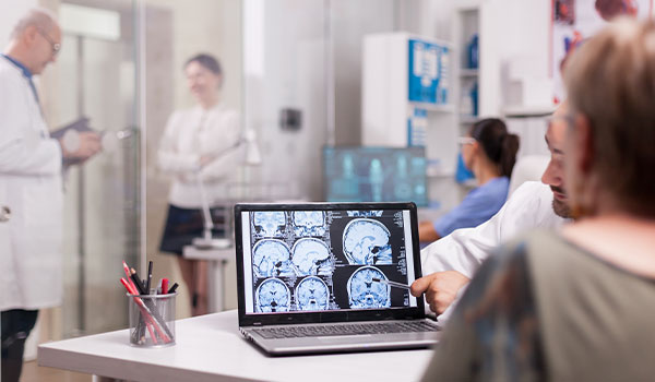
MRI can help identify structural changes, inflammation, certain tumours, disc abnormalities, or areas affecting the spinal cord or nerves.
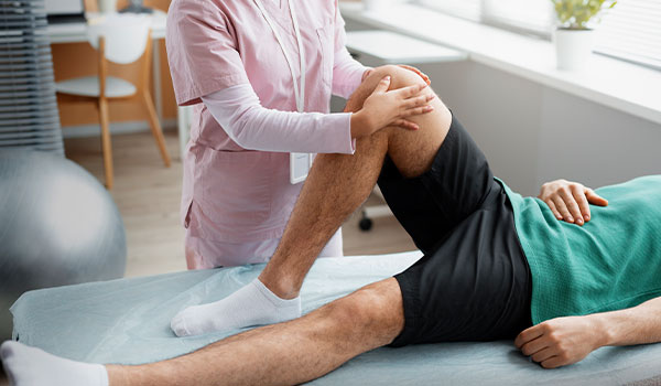
it is recommended after sports injuries, falls or persistent joint symptoms when a doctor needs to assess structures such as ligaments, tendons, cartilage or menisci.

it is used to evaluate structures such as the liver, kidneys, pancreas, reproductive organs, bladder, and surrounding tissues.
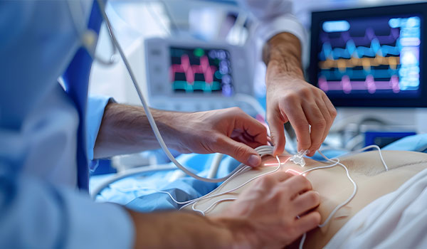
MR angiography can be used to obtain images of blood vessels. It helps doctors assess the structure of blood vessels and investigate abnormalities affecting blood flow or vessel anatomy.
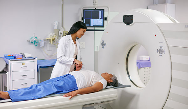
it may be used when doctors need to assess a suspected tumor, determine its location and relationship with nearby structures, or evaluate changes over time. In some cases, contrast-enhanced MRI may provide additional information about abnormal tissue.

it can be used after a condition has already been diagnosed. Repeat scans allow doctors to compare current images with earlier examinations. This is done to evaluate whether a condition has remained stable, changed, or responded to the treatment.
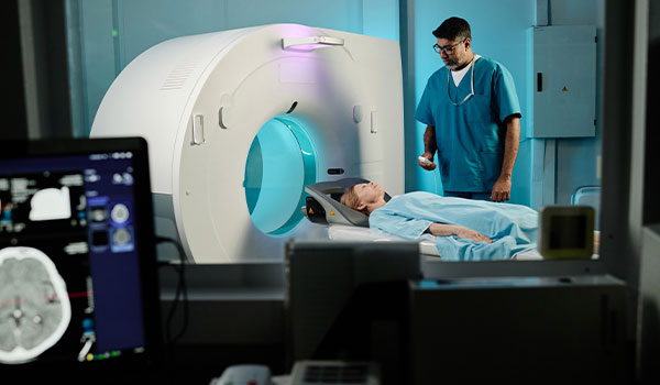
at times, an X-ray, CT scan, or ultrasound does not provide all the information needed to answer a specific clinical question. In such situations, an MRI may be recommended to obtain more detailed images of specific soft tissues and internal structures.
Different MRI examinations are designed for different areas of the body. The choice depends on the suspected condition and the information required by the referring doctor. Below is a list of different magnetic resonance imaging in Siliguri –
| Type of MRI | Why is it used |
|---|---|
| MRI Brain | To investigate conditions such as stroke, brain tumours, certain infections, epilepsy related abnormalities, multiple sclerosis, and other neurological conditions. |
| MRI Spine | When detailed information is required about disc abnormalities, spinal cord changes, nerve compression, certain tumors, inflammation, or other structural problems. |
| MRI knee/joint | It helps assess problems involving ligaments, tendons, cartilage, muscles, menisci, and other soft tissues. |
| MRI abdomen | To evaluate selected abdominal organs and tissues, including liver, kidneys, spleen, pancreas, bowel, and adrenal glands. |
| MRI pelvis | It may be used to evaluate the bladder, reproductive organs, and other pelvic tissues. |
| MR angiography (MRA) | It is used to evaluate blood vessels. It provides information about blood vessel anatomy and some vascular abnormalities |
| Magnetic Resonance Cholangiopancreatography (MRCP) | This technique is used to obtain images of the bile duct and pancreatic duct. |
| MRI with contrast | It makes tissues and blood vessels more conspicuous on MRI images. It provides information that is not visible in non-contrast images. |
An MRI is a non-invasive procedure that takes place in simple steps. Before the scan begins, the technologist checks the medical and implant history to ensure that the examination is performed safely. During the MRI diagnostic test in Siliguri , the patient needs to remain still while the MRI machine captures images of the required body area.
the MRI specialist asks about metal objects, implants, medical devices, previous surgeries, and other relevant medical information.
the patient may be asked to change into a gown and remove items such as jewellery, watches, belts, or anything else containing metal.
a special device called a coil is placed near the area being examined. The examination table moves into the MRI scanner, where images are captured.
if contrast is required, it is administered intravenously. Contrast is not required for every MRI.
The machine produces loud knocking, tapping, or buzzing sounds during scanning. The patient needs to remain as still as possible.
The exact instructions for preparation depend on the body part being examined, type of MRI, whether contrast is required, and the imaging centre’s protocol. Following the instructions provided by the doctor can ensure that the MRI diagnostic imaging in Siliguri is performed safely and produces good quality images. Before the MRI, the patient must keep the following points in mind –
the team must be made aware of any pacemaker, cochlear implant, neurostimulator, or any other metallic device.
if the patient has undergone surgery involving metal devices or implants, the details must be provided to the MRI team.
this information is crucial to decide whether contrast is appropriate.
if the patient is pregnant or thinks that she is pregnant, the radiology team can discuss the appropriate procedure.
if the doctor has instructed the patient to avoid eating or drinking for a specific period, then the patient must adhere to them.
before entering the MRI room, the individual should remove jewellery, watches, belts, and hair accessories containing metal and other electronic devices.
Both MRI and CT scans are advanced imaging tests used to create images of the body. However, they use different technologies and provide different types of information. Understanding the difference between the two examinations can help patients know why a doctor may recommend one test over the other. For patients looking for an MRI test in Siliguri, it is important to know that an MRI is an advanced version of a CT scan.
| Feature | MRI | CT scan |
|---|---|---|
| Imaging method | Magnetic field and radio waves | X-rays and computer processing |
| Ionising radiation | No | Yes |
| Soft-tissue detail | Excellent | Good, depending on the body part |
| Bone assessment | Used for selected conditions | Very useful |
| Scan speed | Slower | Faster |
| Emergency imaging | May not be suitable for every emergency | Frequently used when rapid imaging is needed |
| Contrast | Gadolinium | Iodine based |
The cost of MRI imaging in Siliguri may range between ₹3500 – ₹35000+. The cost varies depending on the body part being scanned, whether contrast is being used, and the type of MRI protocol being used. The table below lists the approximate prices of MRI –
| MRI test | Approximate cost |
|---|---|
| MRI brain, plain | ₹3500 –₹7000 |
| MRI brain with contrast | ₹6000 –₹8000 |
| MRI spine | ₹2500 –₹7500 |
| MRI knee/joint | ₹3500 –₹7000 |
| MRI abdomen | ₹6000 –₹8000 |
| MRI pelvis | ₹5000 –₹8000 |
| MRCP (Magnetic resonance cholangiopancreatography) | ₹Around 7000 |
| MR angiography | ₹5000 –₹8000 |
| MRI whole body | ₹20,000 –₹35000+ |
Note - Magnetic resonance cholangiopancreatography shows bile ducts and gallbladder, pancreas, and pancreatic duct.
MR angiography provides pictures of blood vessels and blood flow.
Disclaimer – These are indicative ranges, not fixed rates. Patients should confirm the latest price, contrast charges, and reporting charges with the diagnostic centre before booking.
The goal of MRI is not just to produce images. It is to provide clinically useful information that can help understand a patient’s condition. The doctor can then plan the further course of action. This is why patients choose Dr. Yugal Goyal for an MRI examination in Siliguri. Here are more reasons why he is the first choice for patients –
Dr. Yugal Goyal holds MBBS and MD in radiodiagnosis with specialised training in diagnostic imaging.
Dr. Goyal evaluates MRI findings in relation to the patient’s symptoms, medical history, and clinical concerns, ensuring that the imaging assessment is relevant to the reason for the scan.
Dr. Goyal can review MRI images remotely, thus supporting timely interpretation and reporting when in-person radiology consultation is not required.
Dr. Yugal Goyal also offers a range of image-guided procedures, including FNAC, percutaneous drainage, nephrostomy, biliary drainage and PTBD stenting, providing patients access to specialised interventional radiology services.
MRI is generally considered a very safe imaging procedure because it does not use ionising radiation. However, it cannot be described as completely risk-free. Due to the strong magnetic field, certain metal objects, implants, and medical devices need to be carefully checked before the scan.
In most cases, you can eat or drink normally before the MRI scan. However, for an abdominal or pelvic MRI, the doctor may ask you to avoid eating for a specified period.
Yes, MRI can show certain types of nerve or nerve-related damage. This happens particularly when the problem involves the spinal cord, nerve roots, or surrounding structures. MRI can provide detailed images of soft tissues and can help identify abnormalities that may affect nerves.
An MRI test in Siliguri usually takes 30 to 60 minutes. The exact duration may vary depending on the body parts being examined and the type of MRI protocol required. Simple MRIs may take 20-30 minutes while more detailed scans may take 45-60 minutes.
An MRI examination is generally painless because there is no cutting or surgery involved. The patient may experience some noise. Other than that, there is no other pain associated with MRI.
No, not all MRI scans require contrast. Many MRI examinations can provide detailed diagnostic information without using any contrast. Whether contrast is needed depends on the body part being examined, the suspected condition, and the information the doctor needs.
For an MRI, choose comfortable, loose-fitting clothing without metal components. Depending on the scan and the imaging centre’s policy, you may be asked to change into the hospital gown.
Yes, having a metal implant does not automatically disqualify you for an MRI. Most modern implants are designed to be MRI-safe or MRI conditional. This means that they can be scanned under specific conditions. However, the exact type, material, location, and model of the implant must be checked before the examination.
What Patients Say
Clear & Reassuring
The entire process was explained clearly, and I felt comfortable asking questions. The consultation was professional, calm and reassuring.

Amit Sharma, Siliguri
Professional Experience
Very professional approach and detailed attention to the case. I appreciated how patiently everything was explained before proceeding.

Priya Das, Siliguri
Thorough Consultation
Dr. Goyal took the time to understand the concern and explain the findings in simple language. A very reassuring experience.

Rahul Agarwal, Jalpaiguri
Patient & Approachable
The consultation was handled with patience and care. Dr. Goyal explained the medical details in a way that was easy to understand.

Sneha Roy, Malbazar
Sanjay Gupta
From the initial consultation to understanding the imaging findings, everything was handled in a clear and professional manner.

Smooth Experience, Siliguri
Copyrights © 2026, Dr. Yugal Goyal.
