
A kidney ultrasound is a non-invasive imaging test used to examine the kidneys and nearby structures, such as the urinary bladder, if recommended by the doctor. It is also called renal ultrasound or renal sonography. The scan is recommended when symptoms, urine findings, blood tests, or an existing kidney condition require further evaluation. For patients looking for kidney ultrasound in Siliguri, Dr. Yugal Goyal provides one of the most precise diagnostic imaging services. His longstanding experience in radiology reinforces people's trust in his medical acumen.
Renal ultrasound is commonly used to assess kidney size, structure, urinary obstruction, stones, and other abnormalities. It gives the healthcare provider information about the health of the kidneys and helps them diagnose certain diseases and conditions.
According to Cleveland Clinic, a kidney or renal ultrasound is an imaging test that allows the healthcare provider to look at the kidneys and the bladder. It uses a transducer that is moved across the kidneys.
It sends out ultrasound waves at an extremely high frequency. The ultrasound waves move through the body to the organs and structures. The organs reflect the sound waves, which are intercepted by the transducer. They are then converted by the computer into images of the tissues and organs being examined.
A kidney ultrasound in Siliguri can help assess:
A kidney ultrasound can provide detailed images of the kidneys and parts of the urinary system. It can help doctors assess the size, shape, position, and structural appearance of the kidneys and identify several abnormalities that may require further evaluation or treatment. Depending on the type of ultrasound and the symptoms of the patient, a kidney ultrasound scan in Siliguri can detect -

it can detect multiple stones along with their size and approximate location. It can also show changes caused by a stone blocking the normal flow of urine. However, many small stones or stones located in certain parts of the urinary tract may not be visible on the ultrasound.

hydronephrosis is the swelling of the kidney's collecting system because urine is not draining normally. A kidney ultrasound can show this and may help doctors understand possible causes, such as a kidney stone or another obstruction in the urinary tract.

fluid-filled structures called kidney cysts can be detected through kidney ultrasound. This may help doctors determine whether it is a simple cyst or requires further assessment.
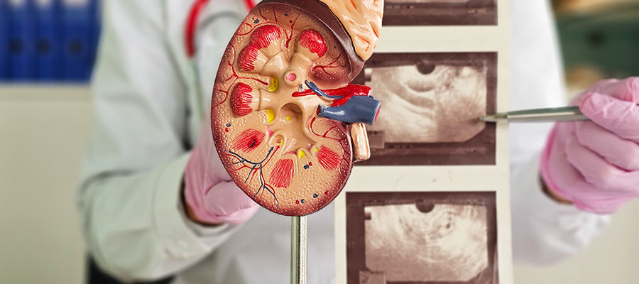
the scan can measure the size of each kidney and examine its overall shape and appearance. Differences in kidney size or structural changes may provide useful information when evaluating certain chronic kidney or urinary conditions.

abnormalities affecting the structure or position of the kidneys can be identified through ultrasound. These findings may be congenital (since birth) or develop over time.

ultrasound may assess a bladder's appearance and the amount of urine remaining after urination (post-void residual urine). This is useful when urinary retention or other urinary problems are suspected.
Recent research also suggests that adding colour Doppler to conventional ultrasound may improve the detection of ureteral stones, especially smaller stones.
| Stone Size | Detection with 2D Ultrasound | Detection with Colour Doppler |
|---|---|---|
| Less than 6mm | 62.50% | 95.83% |
| 6 to 8 mm | 72.73% | 87.88% |

Preparing for a kidney ultrasound is usually simple. However, the exact instructions depend on what needs to be scanned. A routine renal USG in Siliguri requires following the diagnostic centre's instructions, keeping a full bladder if asked to, wearing comfortable clothing for easy access, fasting before the scan if prescribed, and bringing previous reports, etc. These steps have been described below in detail -
before the appointment, confirm the preparation requirements with the diagnostic centre. The instructions may vary depending on the patient's symptoms and the suspected reasons.
if the patient is asked to keep a full bladder, then they may drink water and avoid urinating until the examination is completed. This helps in getting clear images of the tissues and organs.
generally, a renal ultrasound does not require fasting. However, if the doctor has recommended a whole abdominal ultrasound along with the kidney examination, the patient may be asked to avoid food before the scan. This is because gas may interfere with clear images.
wear loose clothing that provides easy access to the abdomen and sides of the body. During the examination, you may need to expose the area being scanned to the ultrasound probe.
inform the radiology team about the reason for the examination, such as flank pain, blood in the urine, urinary difficulties, or kidney stones. This information can help ensure that the scan is performed with appropriate clinical context.
During a kidney ultrasound, the patient is asked to lie on their back or change position during the scan. The sonographer applies ultrasound gel on the abdomen or sides of the body. A transducer is then moved over the required area.
Images of the kidneys are obtained from different angles to clearly assess their appearance. The patient may be asked to hold their breath briefly or change position to obtain clearer images. If the bladder is included in the kidney scan in Siliguri, the radiologist may examine it before and, when clinically required, after the patient urinates. The entire procedure is painless but may include some discomfort or pressure when the transducer is applied, particularly when detailed images are needed.
Generally, there is no recovery required after the ultrasound. As soon as the examination is completed, the patient can return to their normal activities, unless the doctor has advised otherwise.
The images obtained through the kidney ultrasound in Siliguri are then examined by a qualified radiologist. The findings are documented in a report, which is then shared with the patient or referring doctor, as per the centre's reporting process. If the ultrasound shows an abnormality, the doctor may recommend further tests or treatment based on the findings.

The cost of kidney ultrasound can range between ₹600- ₹1500 and more. It may vary depending on factors such as the choice of diagnostic centre, the type of examination, the organs included, and whether additional Doppler imaging is required. The table below lists the prices of kidney ultrasound in Siliguri -
| Ultrasound Examination | Indicative Cost in Siliguri |
|---|---|
| Kidney Ultrasound | ₹600-₹1000 |
| Kidney and Urinary Bladder Ultrasound | ₹700-₹1200 |
| Whole abdomen ultrasound including kidneys | ₹850-₹1500 |
Note: These are indicative ranges for guidance, not fixed prices. The actual cost can vary by diagnostic centre, equipment, reporting requirements, and the exact scan prescribed. Patients should confirm the current fee and preparation requirements with the diagnostic centre before the examination.
For a kidney ultrasound, appropriate imaging technique and accurate interpretation are important because ultrasound findings are understood along with the patient's symptoms and medical history. These features can be found at Dr. Yugal Goyal's ultrasound centre. In addition to these features, he is also the first choice among patients for a kidney USG scan in Siliguri due to the following reasons -
he holds an MBBS and MD in Radiodiagnosis, along with years of experience in kidney ultrasound. This fortifies the trust of patients in his expert ultrasound skills.
Dr. Goyal is one of the very few doctors to provide the service of remote kidney ultrasound interpretation. This helps patients even from locations beyond Siliguri.
Patients receive a clear and detailed interpretation of the ultrasound report. Timely reporting helps patients and physicians move forward with treatment, thereby reducing unnecessary delays.
He uses state-of-the-art imaging technology that provides exceptionally clear images. This prevents the need for a rescan, thus saving time for the patient.
A kidney ultrasound, also called a renal ultrasound, is a non-invasive imaging test that uses high-frequency sound waves to create images of the kidneys. It helps doctors assess the size, shape, and structure of the kidneys and look for conditions such as kidney stones, cysts, swelling of the kidney, or signs of urinary obstruction. It does not use X-rays or ionising radiation.
Yes, a kidney ultrasound is generally considered a safe and painless imaging test. It does not use radiation and usually does not have any significant side effects. Patients may experience mild pressure or discomfort when the transducer is being moved over the skin.
This depends on whether the ultrasound involves only the urinary bladder or kidneys. In the former case, you may be asked to drink water and avoid urinating. This is to keep the bladder full, which provides clear images of the tissues and organs. In the latter case, an ultrasound involving only the kidneys may not require this step. However, you must try to adhere to the instructions of the healthcare provider.
Yes, a kidney ultrasound in Siliguri can detect kidney stones. It may also show hydronephrosis (swelling of the kidney) when a stone is blocking urine flow. However, not every stone can be clearly seen on the ultrasound.
A kidney ultrasound can show structural changes, such as changes in kidney size, appearance, or the presence of cysts or obstruction. However, it cannot determine kidney function by itself. To evaluate kidney disease, blood tests, urine tests, and medical history, etc. are required.
A routine kidney ultrasound generally takes around 15-30 minutes. However, the exact time may vary depending on the type of examination and whether the bladder or some other abdominal organs also need to be examined.
Yes, you can have a kidney ultrasound during pregnancy. This is because ultrasound uses high-frequency sound waves that are non-invasive. Thus, it is a safe procedure. It does not use X-rays (ionising radiation). However, the examination must be performed based on clinical need and medical advice.
A kidney-only ultrasound does not require fasting. However, preparation may differ if the examination is combined with a whole-abdomen ultrasound. In such cases, the diagnostic centre may recommend fasting for a specific period. Patients need to follow the instructions of the healthcare provider.
What Patients Say
Clear & Reassuring
The entire process was explained clearly, and I felt comfortable asking questions. The consultation was professional, calm and reassuring.

Amit Sharma, Siliguri
Professional Experience
Very professional approach and detailed attention to the case. I appreciated how patiently everything was explained before proceeding.

Priya Das, Siliguri
Thorough Consultation
Dr. Goyal took the time to understand the concern and explain the findings in simple language. A very reassuring experience.

Rahul Agarwal, Jalpaiguri
Patient & Approachable
The consultation was handled with patience and care. Dr. Goyal explained the medical details in a way that was easy to understand.

Sneha Roy, Malbazar
Sanjay Gupta
From the initial consultation to understanding the imaging findings, everything was handled in a clear and professional manner.

Smooth Experience, Siliguri
Copyrights © 2026, Dr. Yugal Goyal.
