
A pelvic ultrasound is a commonly used imaging test that creates real-time images of structures in the lower abdomen and pelvis using high-frequency sound waves. It is a non-invasive examination and does not use ionising radiation. For patients looking for a pelvic ultrasound scan, Dr. Yugal Goyal provides the advanced pelvic ultrasound in Siliguri. His extensive experience in ultrasound services enables him to diagnose complex medical issues precisely.
Depending on the patient’s symptoms, age, sex, and clinical indication, different ultrasound approaches may be used to examine specific pelvic structures. Thus, understanding what the scan examines, how to prepare, how it is performed, and what can affect its cost can make the process easier.
A Pelvic ultrasound is an imaging exam that uses sound waves to create pictures of organs inside the body. These pictures are called sonograms. A pelvic ultrasound looks at the organs in your pelvic area between the abdomen(belly) and the legs. The pelvic organs include the bladder, fallopian tubes ( which carry eggs between the ovaries and the uterus), ovaries, prostate, rectum, uterus, and vagina.
The exact structures that are to be examined depend on the type of pelvic ultrasound prescribed by the doctor. It may be recommended to investigate symptoms, assess a known condition, monitor certain findings, or provide additional information alongside other clinical examinations and tests.
Pelvic ultrasound can be performed using different approaches. The most suitable method depends on the type of ultrasound prescribed and the structures to be assessed. The different types of pelvic sonography in Siliguri are described below –

the transducer is moved over the lower abdomen after applying gel to the skin. Patients are usually instructed to keep a full bladder(drink water and avoid urinating) to get clear images. This provides an overview of pelvic structures and is applicable for both males and females.

it uses a specially designed transducer that is gently introduced into the vagina to obtain closer images of the pelvic regions. As opposed to Transabdominal pelvic ultrasound, the bladder is emptied for TVS. A protective cover and lubricant are used on the transducer. The images are obtained from different positions.

in this, the transducer is introduced through the rectum. It is used in selected male patients when the doctor requires detailed imaging of structures such as the prostate.

Not every pelvic ultrasound examines every structure. The areas to be examined depend on what the doctor has recommended and the patient’s clinical indication.
it assesses the size, shape, position, and overall appearance of the uterus. It is performed to help identify structural changes such as fibroids, polyps, or other abnormalities that require further evaluation.
this is the inner lining of the uterus. Ultrasound is used to assess its thickness and appearance. This is usually performed to evaluate abnormal uterine bleeding, menstrual concerns, infertility, or changes after menopause.
to assess the size, location, and appearance of the ovaries, ultrasound is recommended. It helps in identifying ovarian follicles, cysts, and other structural findings. It may also be used to monitor ovarian follicles when clinically required.
the cervix is the lower part of the uterus that connects with the vagina. Its appearance and certain structural changes may be assessed as part of the pelvic examination.
these areas are situated beside the uterus and include structures such as the ovaries and the fallopian tubes. Ultrasound is used to assess these areas for findings such as cystic or solid masses.
ultrasound assesses the urinary bladder for its general appearance, wall changes, bladder contents, or other relevant findings.
depending on the patient’s symptoms and the examination requested, the sonographer or radiologist may assess other visible structures within the pelvic region. The scope of the examination varies from patient to patient and depends on the clinical question being investigated.

Pelvic ultrasound is used to assess how well the bladder empties by measuring post-void residual urine (when specifically requested). At other times, bladder assessment is done to investigate symptoms such as difficulty urinating, frequent urination, or a feeling of incomplete bladder emptying.

Ultrasound is performed to obtain information about the prostate’s size, shape, and general appearance. Prostate imaging is prescribed by the doctor to evaluate urinary symptoms, an enlarged prostate, or other prostate-related concerns.

They are glands located behind the urinary bladder that contribute fluid to the semen. Ultrasound can assess their general appearance and identify structural changes.

Depending on the clinical requirements, an ultrasound may also examine other structures within the male pelvis. The exact areas examined can vary according to the patient’s symptoms, medical history, and the ultrasound technique being used.
A doctor may recommend a pelvic ultrasound when symptoms, findings, medical history, or follow-up requirements indicate that imaging could provide useful information. Common reasons why doctors prescribe pelvic ultrasound in Siliguri are -
unexplained or persistent pain in the lower abdomen or pelvic region may require imaging to look for any structural causes.
ultrasound may be recommended for unusual, irregular, or heavy bleeding, including cases of bleeding after menopause.
when a doctor suspects a mass in the pelvic region, ultrasound is used to obtain images of relevant structures.
pelvic ultrasound may be recommended when a woman experiences changes in her menstrual cycle. This may include irregular or missed periods, heavy or prolonged bleeding, painful periods, suspected ovarian abnormalities, or suspected uterine abnormalities.
ultrasound allows clinicians to assess pelvic reproductive structures and, if required, monitor specific findings during treatment.
The procedure of pelvic ultrasound varies depending on the type of ultrasound.
Preparation for a pelvic ultrasound depends on the type of examination the patient has been asked to undergo. For some transabdominal examinations, the patient may be asked to drink water and avoid urinating until the ultrasound is complete. A full bladder improves the images obtained.
For a transvaginal ultrasound, the bladder is emptied before the examination. Patients are advised to wear comfortable clothing to allow easy access to the area being examined. However, depending on the examination, patients are sometimes asked to change into a gown.
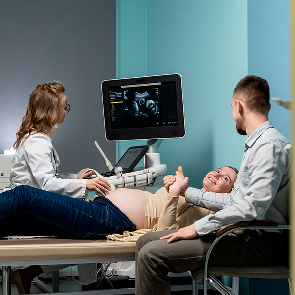
The cost of pelvic sonography in Siliguri is usually between ₹500–₹1900 depending on the diagnostic centre and the type of facility.
| Types of Ultrasound | Approximate Cost |
|---|---|
| Transabdominal ultrasound | ₹800–₹1500 |
| Transvaginal ultrasound | ₹1000–₹2000 |
| Transrectal ultrasound | ₹1500–₹2500 |
| Pelvic ultrasound with Doppler | ₹1500–₹3000 |
| Pregnancy ultrasound | ₹600–₹1500 |
Disclaimer : These are indicative ranges and should not be treated as fixed prices. The actual cost of an ultrasound in Siliguri may vary depending on the type of examination, whether Doppler or additional imaging is required, the diagnostic centre, and the complexity of the examination. Patients should confirm the current price with the centre before undergoing the scan.
When choosing a specialist for pelvic ultrasound, the experience and qualifications of the doctor interpreting the images are important. Dr. Yugal Goyal is a consultant radiologist with MBBS and MD in Radiodiagnosis. With expertise in diagnostic imaging and ultrasonography, he assesses pelvic structures based on the patient’s symptoms and the specific clinical requirement. Here are some other reasons why he is the top choice for pelvic ultrasound in Siliguri -
Dr. Yugal Goyal holds an MBBS and an MD in Radiodiagnosis. This reinforces the trust of patients in his exceptional medical abilities.
He has extensive experience in diagnostic imaging, particularly ultrasonography. Thus, he can diagnose complex pelvic issues with ease.
He has a wide array of Image-Guided procedures, including biopsies, FNAC, percutaneous drainage, nephrostomy, PTBD stenting, and more. Thus providing a one-stop solution for patients.
with high-definition imaging technology, the ultrasound images obtained are extremely clear. This prevents the need to go for a rescan, thus saving the patient's time.
his compassionate care has been a constant feedback from patients. He listens patiently and explains the diagnosis in detail, thus preventing any sort of confusion.
No, pelvic ultrasound and transvaginal ultrasound, even though related, are not the same. Pelvic ultrasound is a wider term for ultrasound of the pelvic region. However, a transvaginal ultrasound is a type of pelvic ultrasound in which a special transducer is gently introduced into the vagina to obtain closer and more detailed images of nearby structures.
A pelvic ultrasound is not generally painful. However, some patients may experience mild pressure or discomfort depending on the type of ultrasound. In a transabdominal ultrasound, the patient may feel some pressure, especially when the bladder is full. In a transvaginal ultrasound, the transducer may cause mild pressure or discomfort, especially if the pelvic area is sensitive. In transrectal ultrasound, the transducer is introduced into th rectum and may cause temporary pressure or discomfort.
A pelvic ultrasound usually takes around 15-30 minutes. However, the exact duration can vary depending on the type of examination and the structures that need to be assessed. The actual scanning time is generally shorter than the total time spent at the diagnostic centre, as preparation and reporting may require additional time.
Yes, you can eat normally after a pelvic ultrasound in most cases. Unlike some abdominal ultrasound scans, a normal pelvic ultrasound does not require fasting. However, preparation may vary depending on the type of pelvic ultrasound.
Yes, a pelvic ultrasound can detect an early pregnancy, but how early it can be seen depends on the gestational age, the type of ultrasound used, and individual factors. For this, a transvaginal ultrasound is usually performed because it can provide early and detailed imaging of a pregnancy.
Pelvic Ultrasound is considered a safe and non-invasive imaging examination. It uses high-frequency sound waves to create images and does not use ionising radiation. Most people do not experience any significant side effects. There might be temporary pressure or discomfort from the transducer, particularly with a full bladder or when it is introduced into the vagina during a transvaginal ultrasound. These issues settle after the examination.
Yes, pelvic ultrasound is generally considered a safe imaging procedure when performed appropriately. It uses high-frequency sound waves instead of ionising radiation, unlike X-Rays and CT scans. Its safety is attested by the fact that it is commonly used during pregnancy to monitor the growing fetus.
Yes, you can have an ultrasound during your period. Menstrual bleeding does not prevent the examination from being performed.
What Patients Say
Clear & Reassuring
The entire process was explained clearly, and I felt comfortable asking questions. The consultation was professional, calm and reassuring.

Amit Sharma, Siliguri
Professional Experience
Very professional approach and detailed attention to the case. I appreciated how patiently everything was explained before proceeding.

Priya Das, Siliguri
Thorough Consultation
Dr. Goyal took the time to understand the concern and explain the findings in simple language. A very reassuring experience.

Rahul Agarwal, Jalpaiguri
Patient & Approachable
The consultation was handled with patience and care. Dr. Goyal explained the medical details in a way that was easy to understand.

Sneha Roy, Malbazar
Sanjay Gupta
From the initial consultation to understanding the imaging findings, everything was handled in a clear and professional manner.

Smooth Experience, Siliguri
Copyrights © 2026, Dr. Yugal Goyal.
